X-Ray Phosphors
X-ray phosphors absorb X-Rays and emit visible light. A common use is as a coating for a scintillation screen with a visible light detector or piece of film. We offer X-ray phosphors, X-ray scintillation cards or screens and X-ray sensitive cameras.
You can see these phosphors in our online store by clicking here.
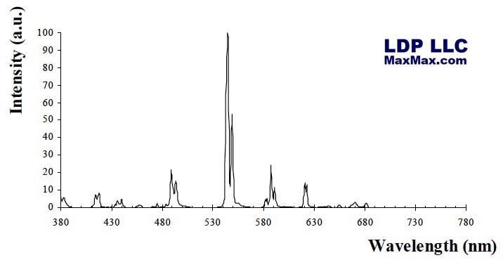
XRay1: Gd2O2S:Tb (P43), green (peak at 545 nm), 1.5 ms decay to 10%, low afterglow, high X-ray absorption, for X-ray, neutrons and gamma, hard X-Rays (>10keV). This phosphor has strong X-ray absorption at the Gd K-edge of 50KeV which is in the midle of the diagnostic X-ray engergy range and a very high light yield around 40,000 photonsMeV. The phosphor crystalizes in a perfect polyhedra which is an important parameter for reaching high spatial resolution.
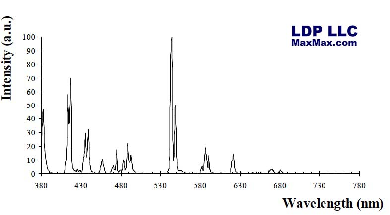
XRay2: Y2O2S:Tb (P45), white (545 nm), 1.5 ms decay, low afterglow, for low-energy X-ray, soft X-Ray, <10keV, This phosphor is typically used for softer X-ray applications such as coating film to convert X-ray energy to visible light. 4.9KeV peak efficiency.
X-rays are at the short wavelength, high energy end of the electromagnetic spectrum. Only gamma rays carry more energy. It is convenient to describe x-rays in terms of the energy they carry, in units of thousands of electron volts (keV). X-rays have energies ranging from less than 1 keV to greater than 100 keV.
Hard x-rays are the highest energy x-rays, while the lower energy x-rays are referred to as soft x-rays. The distinction between hard and soft x-rays is not well defined. Hard x-rays are typically those with energies greater than around 10 keV. More relevant to the distinction are the instruments required to observe them and the physical conditions under which the x-rays are produced.
X-rays do not penetrate the Earth's atmosphere. Therefore they must be observed from a platform launched above most of our atmosphere. The detection of x-rays requires that they interact with a volume of material within the detector, creating free electrons that are ultimately detected as an electric current. Since hard x-rays can penetrate more deeply into a substance than soft x-rays, they require a denser, more massive material to be detected.
X-rays are difficult to reflect and, therefore, image because of their ability to penetrate matter. The wavelengths of x-rays are on the order of or smaller than the size of atoms. As a result they appear less like waves that can be reflected and more like particles (photons) that can effectively pass between the atoms. They penetrate deeply into a material before interacting with an individual atom.
It is possible to produce a mirror to image soft x-rays if the x-rays hit the mirror at small angles to the reflecting surface. This is known as a grazing incidence telescope. This does not work with hard x-rays, however. Obtaining images of hard x-ray sources has required a less direct approach called Fourier-synthesis imaging. This approach uses pairs of filters, or collimators, to obtain information about the source structure on different size scales. The signals obtained from each of the collimators are combined on a computer to construct the image. The image quality improves as the number of collimators, and the number of size scales sampled, increases.
The first x-ray images above 30 keV have been obtained with the Hard X-ray Telescope on the Yohkoh satellite. The figure below compares the hard x-ray image from one flare with white light and soft x-ray images. The hard x-ray image is similar to the white light image. The soft x-rays, however, come from a more extended region than the hard x-rays.

